Dental X-ray
Dental X-rays are an important tool for accurately diagnosing dental problems, planning therapies and monitoring oral health. With the help of quality recordings, we provide our patients with the best possible protection and treatment.
What is a dental X-ray?
Dental X-ray enables accurate diagnosis of teeth, jaws and surrounding structures with the help of X-ray images. It is a tool for early detection of dental problems such as caries, cysts, tumors or abnormalities in tooth development. X-rays are necessary for planning complex procedures such as implants, orthodontic therapies and oral surgery.
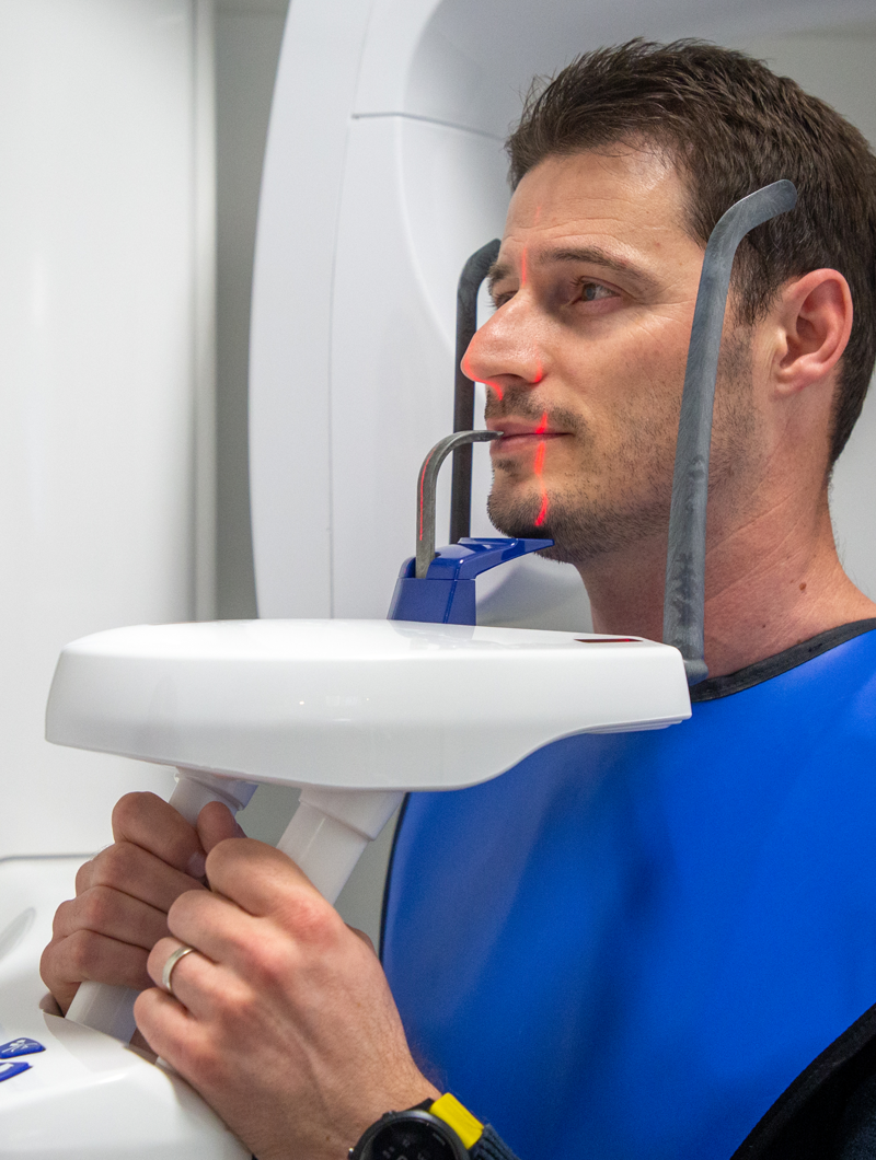
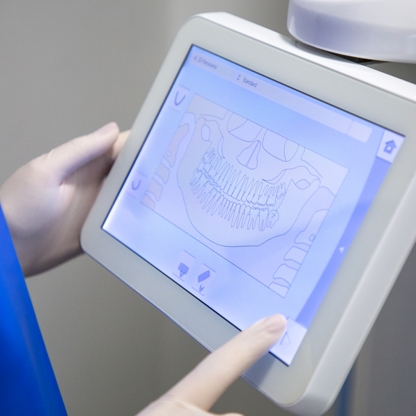
Advanced technologies such as RVG (radiovision graphy) and orthopantomograms enable reduced exposure to radiation, with high-quality images that clearly show the condition of the patient's teeth and jaws.
Who needs a dental X-ray?
A dental X-ray is necessary for patients who need a detailed analysis of the condition of their teeth and jaws. This includes:
Diagnosing caries or other dental problems
Patients who are planning orthodontic treatments or implant procedures
People with complex dental problems who are looking for detailed insight into bone and soft structures
Patients undergoing oral surgery
Process
Frequently asked questions
How much radiation is there during an RVG scan?
The RVG method uses a minimal amount of radiation compared to traditional X-rays, but it provides safe and fast high-quality images. Given the digital format, radiation is significantly reduced compared to classic X-ray films, and patients are exposed to very small amounts of radiation.
Why is an orthopan scan important for my treatment?
An orthopan scan provides a complete picture of your teeth, jaws, temporomandibular joints and sinuses. This is essential for planning orthodontic treatments, as well as for precise monitoring of the condition and prevention of future problems. An orthopan gives your dentist a comprehensive view of your oral health status.
Are there any risks to pregnant women from dental scans?
Pregnant women are usually advised to avoid X-rays, unless necessary for medical reasons. If the scan is necessary, appropriate measures are taken to minimize any radiation exposure. It is always recommended to consult a doctor before making a decision.
What are the advantages of 3D CBCT imaging in dentistry?
3D CBCT imaging provides a detailed view of the three dimensions of the jaw, teeth and surrounding structures. This imaging is particularly useful for implantology procedures because it allows for precise planning of implant placement, assessment of bone quality and analysis of anatomical variations – which helps in the precise performance of the procedure.
How to prepare for orthopan?
Preparation for orthopan is very simple: it is usually necessary to remove jewelry and all metal objects from the head area, as well as clothing that may interfere with the imaging. Patients should follow the dentist's instructions on head positioning and body posture to ensure the image is as clear as possible.